Shoulder
Shoulder Conditions
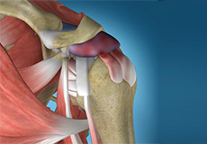
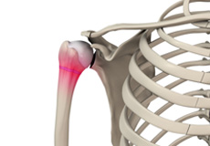
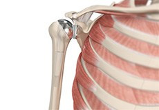
Rotator Cuff Tear
Rotator cuff is the group of tendons in the shoulder joint providing support and enabling wider range of motion. Major injury to these tendons may result in tear of these tendons and the condition is called as rotator cuff tear. It is one of the most common causes of shoulder pain in middle aged adults and older individuals.
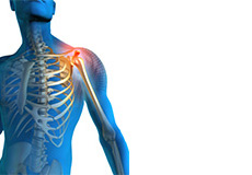
Shoulder Pain
Pain in the shoulder suggests a shoulder injury which is more common in athletes participating in sports such as swimming, tennis, pitching and weightlifting. The injuries are caused due to the over usage or repetitive motion of the arms.
Subluxation
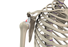
The shoulder is a highly mobile ball and socket joint. The ball of the upper arm bone (humerus) is held in place at the socket (glenoid) of the shoulder blade (scapula) by a group of ligaments.
Shoulder Impingement
Shoulder impingement is the condition of inflammation of the tendons of the shoulder joint. It is one of the most common causes of pain in the adult shoulder.
SLAP Tears
The shoulder joint is a ball and socket joint. A 'ball' at the top of the upper arm bone (the humerus) fits neatly into a 'socket', called the glenoid, which is part of the shoulder blade (scapula). The term SLAP (superior - labrum anterior-posterior) lesion or SLAP tear refers to an injury of the superior labrum of the shoulder.
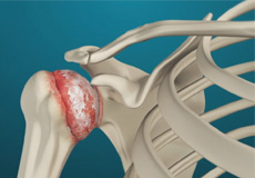
Arthritis of the Shoulder
The term arthritis literally means inflammation of a joint, but is generally used to describe any condition in which there is damage to the cartilage. Damage of the cartilage in the shoulder joint causes shoulder arthritis. Inflammation is the body's natural response to injury. The warning signs that inflammation presents are redness, swelling, heat and pain.
Frozen Shoulder
Frozen shoulder, also called adhesive capsulitis is a condition characterized by pain and loss of motion in shoulder joint. It is more common in older adults aged between 40 and 60 years and is more common in women than men.
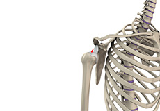
Shoulder Instability
A dislocation occurs when the end of the humerus (the ball portion) partially or completely dislocates from the glenoid (the socket portion) of the shoulder. A partial dislocation is referred to as a subluxation whereas a complete separation is referred to as a dislocation.
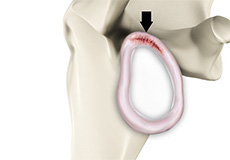
Shoulder Joint Tear
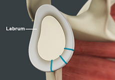
The shoulder joint is a "ball and socket" joint that enables the smooth gliding and thereby the movements of arms. However, it is inherently unstable because of the shallow socket. A soft rim of cartilage, the labrum lines the socket and deepens it so that it accommodates the head of the upper arm bone better.
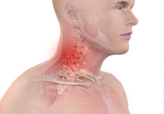
Thoracic Outlet Syndrome
The thoracic outlet is a small passageway leading from the base of the neck to the armpit and arm. This small area contains many blood vessels, nerves and muscle. When this passageway becomes compressed the condition is termed as thoracic outlet syndrome.
Shoulder Separation (AC Dislocation)
Acromioclavicular joint (AC joint) dislocation or shoulder separation is one of the most common injuries of the upper arm. It involves separation of the AC joint and injury to the ligaments that support the joint. The AC joint forms where the clavicle (collarbone) meets the shoulder blade (acromion).
Little League Shoulder
Little league shoulder is an injury to the growth plate of the upper arm bone in the shoulder joint of children. It is caused due to overuse from pitching or throwing, especially in children between the ages of 10 to 15 years. This condition is mostly seen in baseball pitchers, but children in other sports who use improper throwing action are also at risk.
Bicep Tendon Tear at Elbow
The biceps muscle, located in the front of the upper arm allows you to bend the elbow and rotate the arm. Biceps tendons attach the biceps muscle to the bones in the shoulder and in the elbow.
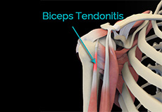
Bicep Tendon Rupture
The biceps tendon is a tough band of connective fibrous tissue that attaches your biceps muscle to the bones in your shoulder on one side and the elbow on the other side.
Burners and Stingers
Burners and stingers are common neck or shoulder injuries characterized by intense burning or stinging pain which can radiate from the neck to the hand. They are caused by sudden movement or a direct blow to the neck resulting in an injury to the brachial plexus.
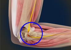
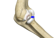
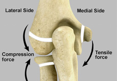
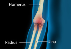
Elbow Dislocation
The elbow is a hinge joint made up of 3 bones - humerus, radius and ulna. The bones are held together by ligaments to provide stability to the joint. Muscles and tendons move the bones around each other and help in performing various activities. Elbow dislocation occurs when the bones that make up the joint are forced out of alignment.
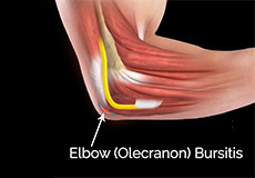
Elbow (Olecranon) Bursitis
The elbow contains a large, curved, pointy bone at the back called the olecranon, which is covered by the olecranon bursa, a small fluid-filled sac that allows smooth movement between the bone and overlying skin. Inflammation of this bursa leads to a condition called olecranon bursitis.
Ulnar Nerve Entrapment
The ulnar nerve travels down the back of the elbow behind the bony bump called the medial epicondyle, and through a passageway called the cubital tunnel. The cubital tunnel is a narrow passageway on the inside of the elbow formed by bone, muscle, and ligaments with the ulnar nerve passing through its center. The roof of the cubital tunnel is covered with a soft tissue called fascia.
Osteochondritis Dissecans
Osteochondritis dissecans is a joint condition in which a piece of cartilage, along with a thin layer of the bone separates from the end of the bone because of inadequate blood supply. The separated fragments are sometimes called "joint mice". These fragments may be localized, or may detach and fall into the joint space causing pain and joint instability.
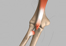
Elbow Sprain
Elbow sprain is an injury to the soft tissues of the elbow. It is caused due to stretching or tearing (partial or full) of the ligaments which support the elbow joint. Ligaments are a group of fibrous tissues that connect one bone to another in the body.
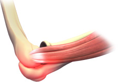
Tennis Elbow
Tennis elbow is the common name used for the elbow condition called lateral epicondylitis. It is an overuse injury that causes inflammation of the tendons that attach to the bony prominence on the outside of the elbow (lateral epicondyle). It is a painful condition occurring from repeated muscle contractions at the forearm that leads to inflammation and micro tears in the tendons that attach to the lateral epicondyle.
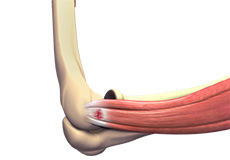
Golfer’s Elbow
Golfer's elbow, also called Medial Epicondylitis, is a painful condition occurring from repeated muscle contractions in the forearm that leads to inflammation and microtears in the tendons that attach to the medial epicondyle. The medial epicondyle is the bony prominence that is felt on the inside of the elbow.
Elbow Injuries
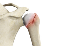
Elbow Fractures: Fracture is a common injury to the elbow. Elbow fractures may result from a fall onto an outstretched wrist, a direct impact to the elbow or a twisting injury. Elbow fractures may cause severe pain, swelling, tenderness and painful movements. If a fracture is suspected, immediate intervention by your doctor is necessary. Surgery is often required if a bony displacement is observed.
Little League Elbow
Little league elbow also called as medial apophysitis, is an overuse condition that occurs when there is overstress or injury to the inside portion of the elbow. It is commonly seen in children involved in sports activities that require repetitive throwing such as baseball.
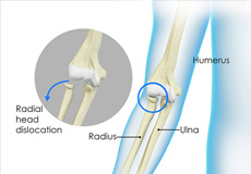
Nursemaid's Elbow
Dislocation of the radius bone present in the elbow is called nursemaid's elbow. This condition is very common among children below 5 years of age as their bones and muscles are still in the developing stage. This condition usually occurs when a child is pulled up too hard by the arm, but can also occur due to a fall or swinging your child by the arms.
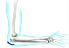
Elbow Pain
The elbow is a hinge joint made up of 3 bones - humerus, radius and ulna. The bones are held together by ligaments to provide stability to the joint. Muscles and tendons move the bones around each other and help in performing various activities.
Fractures/ Injuries
Shoulder injuries most commonly occur in athletes participating in sports such as swimming, tennis, pitching, and weightlifting. The injuries are caused due to the over usage or repetitive motion of the arms.
Shoulder Procedures
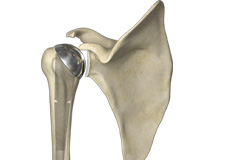
Shoulder Joint Replacement
The shoulder is a highly movable body joint that allows various movements of the arm. It is a ball and socket joint, where the head of the humerus (upper arm bone) articulates with the socket of the scapula (shoulder blade) called the glenoid. The two articulating surfaces of the bones are covered with cartilage, which prevents friction between the moving bones.
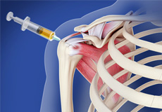
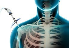
Shoulder Injections
Ultrasound is a common imaging technique that employs high frequency sound waves to create images of organs and other internal structures of the body. These images provide valuable information of underlying pathology of the tissues and assists with diagnosis and planning the treatment of a condition. Ultrasound provides a clear view of the organs, tendons, muscles or joints and any associated disorders.
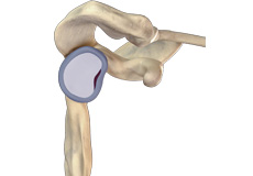
Partial Shoulder Replacement
Partial shoulder replacement, also called shoulder hemiarthroplasty is a surgical procedure during which the upper bone in the arm (humerus) is replaced with a prosthetic metal implant, whereas the other half of the shoulder joint (glenoid or socket) is left intact.

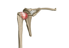
Reverse Shoulder Replacement
Reverse total shoulder replacement, is an advanced surgical technique specifically designed for rotator cuff tear arthropathy, a condition where the patient suffers from both shoulder arthritis and a rotator cuff tear.
Revision Shoulder Replacement
Total shoulder replacement is the replacement of the head of the humerus (upper arm bone) and the glenoid cavity (cavity of the shoulder blade) into which the humerus fits, with artificial prostheses to relieve pain, swelling and stiffness caused due to damage of cartilage at the articulating surfaces.
Minimally Invasive Shoulder Joint Replacement
Shoulder joint replacement is a surgical procedure to replace damaged bone surfaces with artificial components to relieve pain and improve functional ability in the shoulder joint. Shoulder joint replacement can be done by a traditional "open" approach or through a minimally invasive approach.
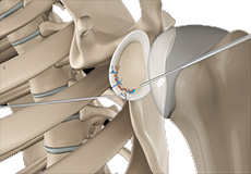
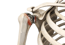
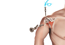
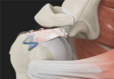
Arthroscopic Bankart Repair
The shoulder joint (glenohumeral joint) is a ball and socket joint, where the head of the upper arm bone (humerus) attaches to the shoulder socket (glenoid cavity). The shoulder socket is extremely shallow and therefore needs additional support to keep the shoulder bones from dislocating.
Shoulder Labrum Reconstruction
The shoulder joint is a ball and socket joint. A 'ball' at the top of the upper arm bone (the humerus) fits neatly into a 'socket', called the glenoid, which is part of the shoulder blade (scapula). The labrum is a ring of fibrous cartilage surrounding the glenoid which helps in stabilizing the shoulder joint.
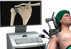
Shoulder Arthroscopy
Arthroscopy is a minimally invasive diagnostic and surgical procedure performed for joint problems. Shoulder arthroscopy is performed using a pencil-sized instrument called an Arthroscope. The arthroscope consists of a light system and camera to project images to a computer screen for your surgeon to view the surgical site.
Arthroscopic Rotator Cuff Repair
Rotator cuff is the group of tendons in the shoulder joint providing support and enabling wider range of motion. Major injury to these tendons may result in tear of these tendons and the condition is called as rotator cuff tear. It is one of the most common causes of shoulder pain in middle aged adults and older individuals.
Superior Capsule Reconstruction
The shoulder joint is stabilized by the joint capsule and rotator cuff. Tears to the rotator cuff can cause severe pain and impairment. When defects in the underlying upper joint capsule add to the instability caused by rotator cuff tears, it cannot be repaired with conventional treatments.

Non Operative / Alternative Options
Alternative options utilize the body’s natural healing mechanism to treat various conditions. Alternative medicine has the potential to renew and repair diseased or damaged tissues and have shown promising results in treatments of various orthopedic conditions.
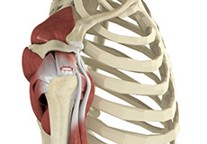
Shoulder Anatomy
The shoulder is the most flexible joint in the body enabling a wide range of movements including, forward flexion, abduction, adduction, external rotation, internal rotation, and 360-degree circumduction.
Thus, the shoulder joint is considered the most insecure joint of the body but the support of ligaments, muscles and tendons function to provide the required stability.
Bones
The shoulder is a ball and socket joint made up of three bones, namely the humerus, scapula, and clavicle.
The end of the humerus or upper arm bone forms the ball of the shoulder joint. An irregular shallow cavity in the scapula called the glenoid cavity forms the socket for the head of the humerus to fit in. The two bones together form the glenohumeral joint, which is the main joint of the shoulder.
The scapula is a flat triangular shaped bone that forms the shoulder blade. It serves as the site of attachment for most of the muscles that provide movement and stability to the joint. The scapula has four bony processes - acromion, spine, coracoid and glenoid cavity. The Acromion and coracoid process serve as places for attachment of the ligaments and tendons.
The clavicle bone or collarbone is an S-shaped bone that connects the scapula to the sternum or breastbone. It forms two joints: the acromioclavicular joint, where it articulates with the acromion process of the scapula, and the sternoclavicular joint where it articulates with the sternum or breast bone. The clavicle also forms a protective covering for important nerves and blood vessels that pass under it from the spine to the arms.
Soft tissues
The ends of all articulating bones are covered by smooth tissue called articular cartilage which allows the bones to slide over each other without friction enabling smooth movement. Articular cartilage reduces pressure and acts as a shock absorber during movement of the shoulder bones.
Extra stability to the glenohumeral joint is provided by the glenoid labrum, a ring of fibrous cartilage that surrounds the glenoid cavity. The glenoid labrum increases the depth and surface area of the glenoid cavity to provide a more secure fit for the half-spherical head of the humerus.
Ligaments
Ligaments are the thick strands of fibers that connect one bone to another. The ligaments of the shoulder joint include
- Coraco-clavicular ligaments: these ligaments connect the collarbone to the shoulder blade at the coracoid process
- Acromio-clavicular ligament: this connects the collarbone to the shoulder blade at the acromion process
- Coraco-acromial ligament: It connects the acromion process to the coracoid process
- Glenohumeral ligaments: A group of 3 ligaments that form a capsule around the shoulder joint, and connect the head of the arm bone to the glenoid cavity of the shoulder blade. The capsule forms a water-tight sac around the joint. Glenohumeral ligaments play a very important role in providing stability to the otherwise unstable shoulder joint by preventing dislocation.
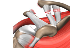
Muscles
The rotator cuff is the main group of muscles in the shoulder joint and is comprised of 4 muscles. The rotator cuff forms a sleeve around the humeral head and glenoid cavity, providing additional stability to the shoulder joint while enabling a wide range of mobility.
The deltoid muscle forms the outer layer of the rotator cuff and is the largest and strongest muscle of the shoulder joint.
Tendons
Tendons are strong tissues that join muscle to bone allowing the muscle to control the movement of the bone or joint. Two important group of tendons in the shoulder joint are the biceps tendons and rotator cuff tendons.
Bicep tendons are the two tendons that join the bicep muscle of the upper arm to the shoulder. They are referred to as the long head and short head of the bicep.
Rotator cuff tendons are a group of four tendons that join the head of the humerus to the deeper muscles of the rotator cuff. These tendons provide more stability and mobility to the shoulder joint.
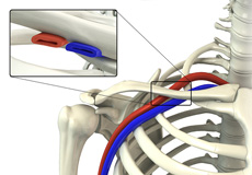
Nerves
Nerves carry messages from the brain to muscles to direct movement (motor nerves) and send information about different sensations such as touch, temperature and pain from the muscles back to the brain (sensory nerves). The nerves of the arm pass through the shoulder joint from the neck.
These nerves form a bundle at the region of the shoulder called the brachial plexus. The main nerves of the brachial plexus are the musculocutaneous, axillary, radial, ulnar and median nerves.
Blood vessels
Blood vessels travel along with the nerves to supply blood to the arms. Oxygenated blood is supplied to the shoulder region by the subclavian artery that runs below the collarbone. As it enters the region of the armpit, it is called the axillary artery and further down the arm, it is called the brachial artery. The main veins carrying de-oxygenated blood back to the heart for purification include:
- Axillary vein: this vein drains into the subclavian vein
- Cephalic vein: this vein is found in the upper arm and branches at the elbow into the forearm region. It drains into the axillary vein.
- Basilic vein: this vein runs opposite the cephalic vein, near the triceps muscle. It drains into the axillary vein.